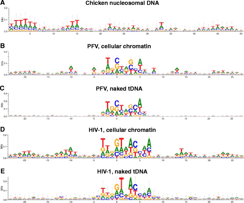
Figure 4.

Sequence logos for PFV and HIV-1 integration sites in nucleosome-free versus chromatinized tDNA. (A) The logo illustrates the average nucleotide sequence of the first 50 nucleotides of center-aligned nucleosomal DNA sequences isolated from chicken erythrocytes [68]. (B) PFV integration sites derived from virus-infected cells [92,93]. (C) Integration sites from recombinant PFV intasomes and deproteinized cellular DNA. (D) HIV-1 integration sites from virus-infected cells [87]. (E) Concerted HIV-1 integration sites from recombinant HIV-1 IN and naked pGEM9Zf(−) plasmid DNA [33].
