Figure 2.

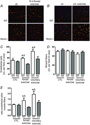
Exercise-induced capillarization is mediated by Nox4
A and B, representative images of gastrocnemius muscle from sedentary (ctl) and exercised (Ex.) wild-type (WT) or Nox4−/− mice, stained for CD31 (green) to show capillaries, as well as laminin (red) to define muscle fibres (see the online version for colours). Sections were made after 10 days (A) and after 4 weeks of voluntary (B) exercise. C–E, statistics. C, endothelial cells per field of view. D, muscle fibre per field of view. E, ratio: endothelial cells per muscle fibre. (n > 5). *P < 0.05 (ctl vs. Ex.); &P < 0.05 (WT vs. Nox4−/−).
