
Fig 4. Br-LPS is released inside PMNs.
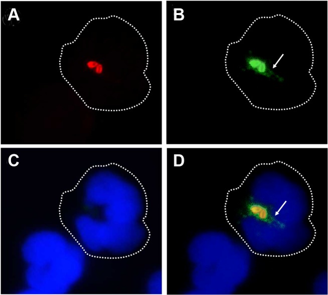
Heparinized blood was incubated with B. abortus-RFP for one hour (MOI 2). Blood smears were fixed, stained with anti-Brucella LPS FITC (green) and mounted with ProLong Gold Antifade Reagent with DAPI. (a) B. abortus-RFP, (b) IgG-FITC anti-Brucella LPS staining, (c) PMN DAPI staining and (d) merged images. Shed Brucella LPS (white arrow) is pointed. Representative PMNs with DAPI-stained nuclei and intracellular B. abortus were photographed under the microscope using the appropriate color filter channel. Images were cut from microscope field, contrasted and saturated using Hue tool to obtain suitable color separation. Images were then merged using Adobe Photoshop 8 program. Microscope images are at 1000 × magnification.