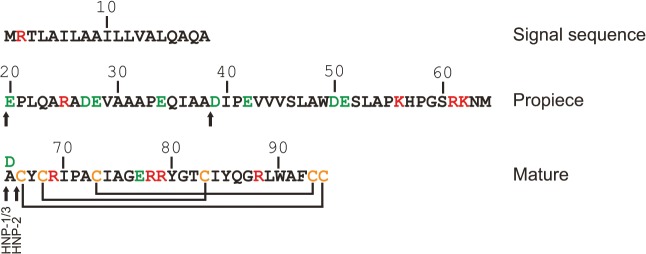
Fig 1. Structure of preproHNP-1-3.

Arrows indicate major sites of proteolytic cleavage. Positively and negatively charged amino acids are indicated in red and green, respectively. Lines indicate the disulphide linkage of cysteines (C; orange). HNP-3 is identical to HNP-1 except for having substituted alanine (A) at position 65 for aspartic acids (D).
