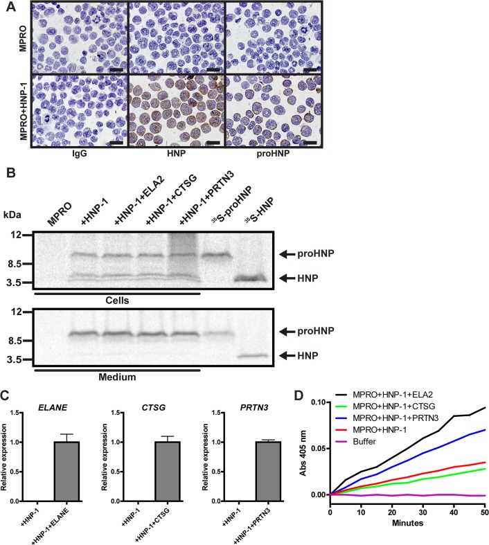
Fig 5. Processing of proHNP-1 by MPRO cells transfected with human serine proteases.
(A) MPRO cells and MPRO cells transfected with HNP-1 were spun onto slides, fixed, permeabilized, and immunocytochemically stained for IgG (top), HNPs (middle), and proHNPs (bottom). Bars represent 20 μm. (B) MPRO-HNP-1 cells were further transfected with the human neutrophil serine proteases neutrophil elastase (ELANE), cathepsin G (CTSG), and proteinase 3 (PRTN3). Cells were pulsed with 35S-methionine/cysteine for 1 hour and chased overnight. Cell lysates and medium were immunoprecipitated with antibodies in the following order: anti-proHNP, and anti-HNP. Immunoprecipitates were pooled and analyzed by 16% SDS-Tricine-PAGE and fluorography using 35S-proHNP and 35S-HNP from the proHNP processing assay as controls. (C) Comparative quantification mRNA for ELANE, CTSG, and PRTN3 was performed by real-time PCR. Figure depicts expression levels relative to cells electroporated human serine proteases. Bars represent means and lines represent standard deviation. (D) Activity of transfected human neutrophil elastase and proteinase 3 was asserted by lysis of transfected MPRO cells followed by spectrophotometry following degradation rate of methoxysuccinyl-Ala-Ala-Pro-Val-P-nitroanilide.

