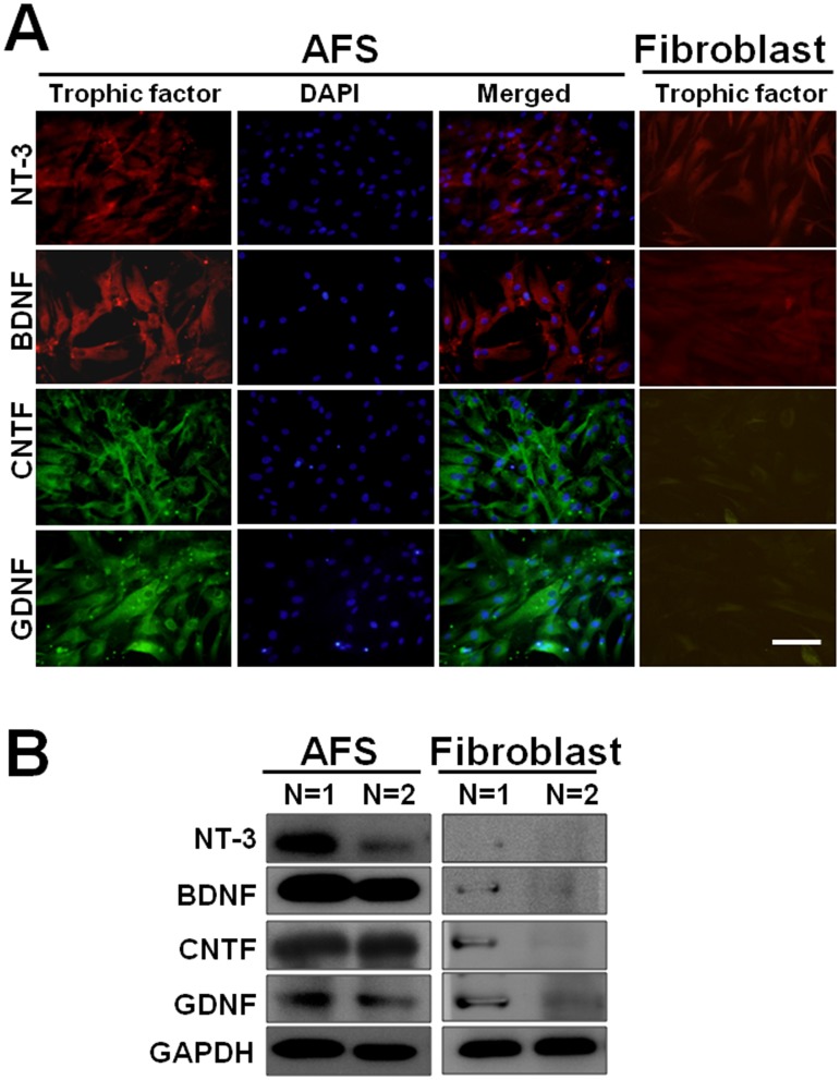
Fig 1. Illustration of neurotrophic factors in AFS.
(A) Positive stating of NT-3, BDNF, CNTF, and GDNF in AFS, counter- staining with DAPI and merged imaging; human fibroblast cells as a negative control (B) The demonstration of expression of NT-3, BDNF, CNTF, and GDNF after Western blot analysis in AFS and human fibroblast cells in two different samples. Bar length = 100 μm.

