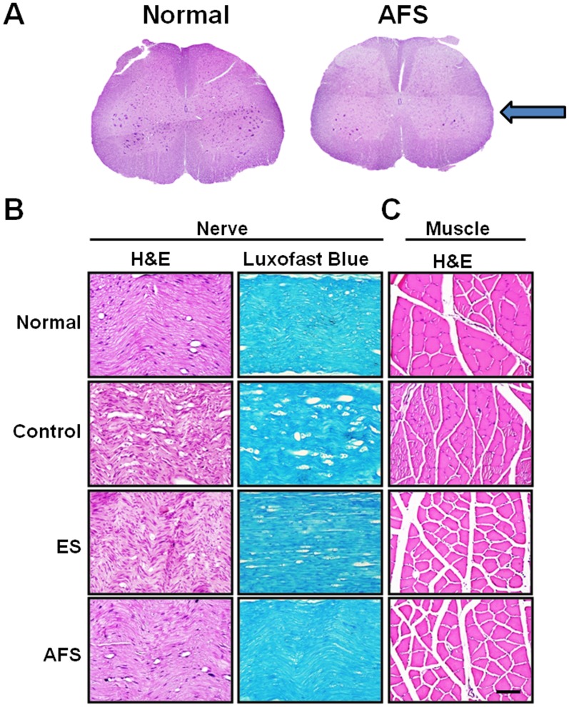
Fig 9. Illustration of morphology alteration of spinal cord, nerve, and muscle 2 months after nerve anastomosis.
(A) morphology alteration in anterior horn cells subjected to AFS treatment (B) morphology and myelination of the distal end of nerve subjected to treatment group (C) morphology alteration in denervated muscle subjected to different treatment group. Bar length = 100 μm; arrow indicated the site of nerve injury.

