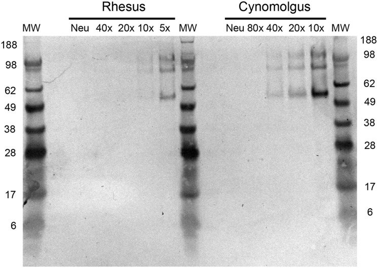
Fig 6. Levels of sialyl-α-2,6-Gal saccharides in trachea tissue samples from rhesus and cynomolgus monkeys.
SNA stained blot of SDS-PAGE separated trachea tissue samples from rhesus and cynomolgus macaques applied in a dilution range of 1:5/1:10/1:20/1:40/1:80 or at a 1:5/1:10 dilution after neuraminidase digestion (Neu). The samples used were a mixture of tissue homogenates from 4 animals. MW = molecular weight marker. Numbers on the left and right side of the gel indicate the molecular weight (kD) of the marker bands.

