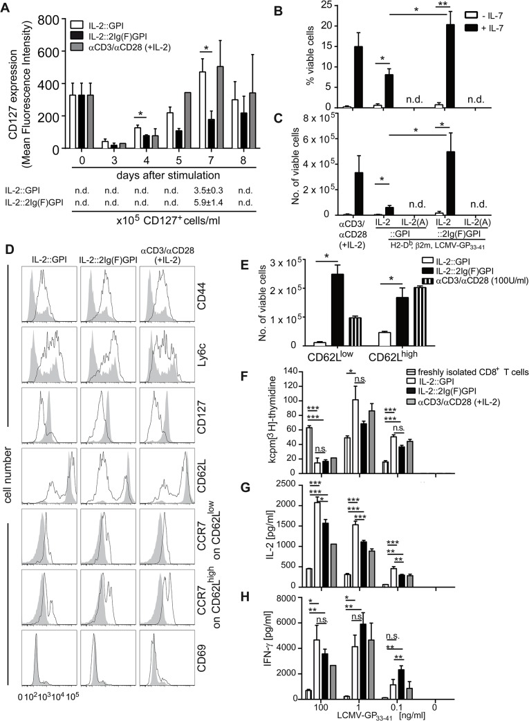
Fig 5. Acquisition of memory-like functions by CD8+ T cells is supported by IL-2 decorated asVNP.
(A) Re-expression kinetics of CD127 on IL-2v asVNP stimulated purified P14 CD8+ TCR Vα2+ T cells. Mean fluorescence intensity of CD127 expression on CD8+ T cells at indicated time points after stimulation with IL-2::GPI, IL-2::2Ig(F)GPI asVNP or αCD3/αCD28 microbeads plus 100 U/ml IL-2. Bottom panel indicates absolute CD127+ cell numbers obtained at day 7. (B) Percentage and (C) absolute numbers of viable (propidium iodide negative) CD8+ T cells upon VNP-mediated primary stimulation for 7 days followed by removal of particles and a 4-day culture ±IL-7 (125 U/ml). n.d, not determinable (T cells did not survive day 7 of primary culture). (D) Flow cytometry analysis showing memory and activation phenotype of naïve (shaded grey histograms) and IL-2v asVNP or αCD3/αCD28 microbeads plus 100 U/ml IL-2 activated cells (black line). (E) Absolute numbers of viable CD8+CD25+CD62Llow and CD8+CD25+CD62Lhigh cells after 11 days of culture (co-culture and resting phase). n.d, not determinable (T cells did not survive day 7 of primary culture). (F—H) Secondary responses of CD8+ T cells upon pre-stimulation with IL-2v. Viable cells (1x10⁴) pre-stimulated as indicated were re-stimulated with LCMV-GP33-41 peptide pulsed or non-pulsed irradiated splenocytes. Freshly isolated CD8+ T cells served as control. (F) Proliferation was determined by [3H]-thymidine incorporation after 48 hours of stimulation. Freshly purified CD8+ T cells served as control. Secreted (G) IL-2 and (H) IFN-γ levels in supernatants of CD8+ T cells during secondary responses. Data are representative (D) or show the summary (A, B, C, E-H) of three (except two for αCD3/αCD28 microbeads plus IL-2) (A), five (except three for αCD3/αCD28 microbeads plus IL-2) (B, C), three (D-H) independent experiments. * p < 0.05, **, p < 0.01, ***, p < 0.001; t-test (A, E), ANOVA and Tukey’s multiple comparison test (B, F-H), Kruskal-Wallis test and Mann-Whitney U-test and Bonferroni correction (C).

