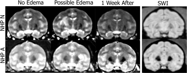
Fig 4. T2-weighted MRI and SWI scans of NHP N and A.
T2-weighted sequences can be used to detect potential edema. The first column shows no atypical hyperintense voxels in the target region. The second column shows atypical hyperintense voxels in the target region. The third column verifies the atypical hyperintense voxels from the previous week were no longer present. The fourth column shows the SWI scans from the day when hyperintense voxels were detected on the T2 scan (acquired the same day as column 2).

