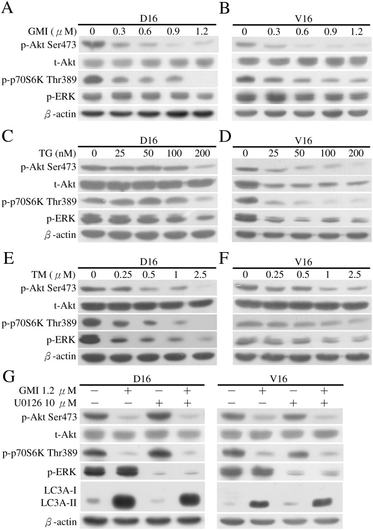
Fig 8. Analyses of Akt, p70S6K and ERK phosphorylation following GMI, TG and TM treatment in MDR sublines.
A549/D16 and (B) A549/V16 sublines were treated with GMI (0.3–1.2 μM) for 48 h and harvested. Equal amounts of total cell lysates were analyzed on Western blot. (C) A549/D16 and (D) A549/V16 sublines were treated with TG (25–200 nM) for 48 h and harvested. (E) A549/D16 and (F) A549/V16 sublines were treated with TM (0.25–2.5 μM) for 48 h and harvested. Protein levels of total Akt (t-Akt), phosphorylated-Akt Ser473 (p-Akt Ser473), phosphorylated-p-70S6K Thr389 (p-p-70S6K Thr389) and phosphorylated-ERK (p-ERK) were determined using the corresponding antibodies. (G) The association of ERK with GMI-induced autophagy was investigated. MDR sublines were pre-treated with U0126 (10 μM) for 1 h followed by GMI (1.2 μM) for 48 h and analysis by Western blotting.

