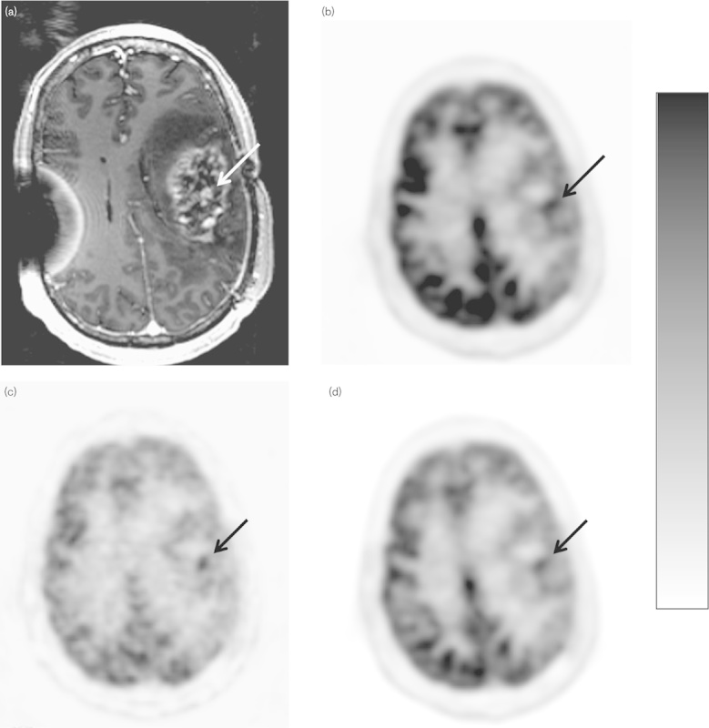
Fig. 4.

Scanning images of a 43-year-old man with high-grade glioma, status postgross total resection and chemoradiation. (a) Postcontrast axial T1-weighted MR image shows enhancement in the left frontoparietal area (arrow). (b) SUVmax axial 18F-FDG PET parametric image, lesion uptake=6.54 (arrow). (c) GMR parametric axial 18F-FDG PET image shows focal uptake [10.21 µmol glucose/min/100 g (arrow)]. (d) Axial SUVgluc 18F-FDG PET image, measured uptake value=4.25 (arrow). For abbreviations, see Fig. 1 legend.
