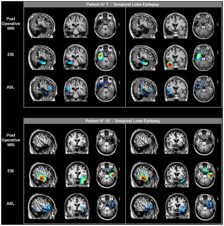
Fig 7. Postoperative imaging in two patients of the new group (patients. nos. 7 and 10).
The presurgical ESI and ASL results are overlaid on the coregistered postoperative MRI scans for each patient. Two different sections are shown for each plane (sagittal, coronal and axial). The rising phase of activity and the statistical results from the one-versus-many analysis are presented for ESI and ASL, respectively.

