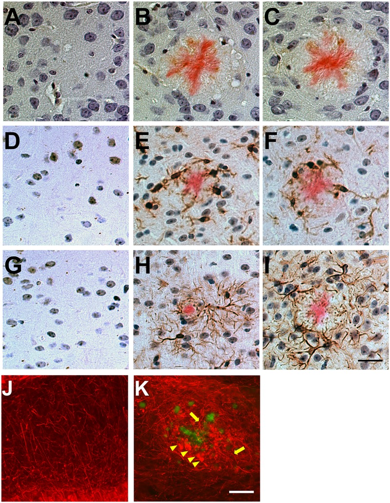
Fig 9. Plaque-associated neuroinflammation and abnormal neuronal architecture in rTg9191 mice.
(A-I) rTg9191 mice show reactive gliosis in the vicinity of dense-core plaques. Brain sections from rTg9191 mice at 24 months of age (B, E, H), their age-matched non-transgenic littermates (A, D, G), and age-matched Tg2576 mice (C, F, I) were stained with antibodies directed against the astroglial marker S100β (A-C), a monoclonal antibody directed against the microglial marker ionized calcium-binding adaptor molecule 1 (Iba1) (D-F), and an antibody directed against the astrocytic marker glial fibrillary acidic protein (GFAP) (G-I). Astrocytes and activated microglial cells and reside near dense-core plaques visualized using Congo red (pink). Scale bar in I, 25 μm, applies to A-I. (J-K) rTg9191 mice exhibited abnormal neuronal architecture around plaques. Thioflavin S (green) was used to visualize plaques and monoclonal antibody SMI-312 was used to visualize axons (red). (J) No plaques were detected in age-matched non-transgenic littermates of rTg9191 mice, and neuronal morphologies were normal. (K) Plaques are surrounded by swollen, dystrophic axons (arrowheads) and curvy, distorted axonal processes (arrows) in brains of rTg9191 mice. Scale bar in K, 50 μm, applies to J and K. Representative photomicrographs show neuroinflammation and neuronal architecture of female mice, and similar results were found in male mice.

