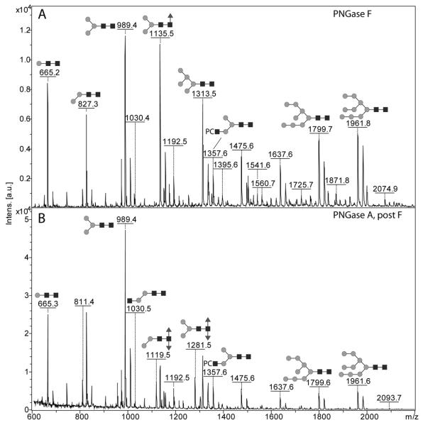
Figure 1. MALDI-TOF mass spectra of the complete N-glycan pools from Pristionchus pacificus.
The N-glycans released by (A) PNGase F or (B) by PNGase A after PNGase F were pyridylaminated (i.e., fluorescently-labelled with 2-aminopyridine) and are annotated with selected major glycan structures; the annotations are for the [M+H]+ forms, but sodiated ions were also detected. The example glycans are depicted according to the symbolic nomenclature of the Consortium for Functional Glycomics; circles, hexose; squares, N- acetylhexosamine; triangles, fucose; PC, phosphorylcholine.

