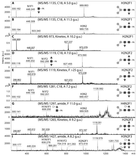
Figure 4. MALDI-TOF MS/MS of pyridylamino-labelled mono-, di- and tri-fucosylated N-glycans.
Examples of MS/MS spectra for glycans monofucosylated on the reducing-terminal GlcNAc (A,B), monofucosylated on the distal GlcNAc (C), difucosylated on the reducing-terminal GlcNAc (D,F), fucosylated on both the reducing-terminal and distal GlcNAc (E), galactosylated on core fucose (G) or trifucosylated on the core (H,I) are shown for glycans identified in various RP-HPLC fractions. Key fragments are annotated with abbreviations of the form HxNyFz where H is hexose, N N-acetylhexosamine, and F fucose or with diagrammatic depictions, in which the α1,3- and α1,6-fucose residues are respectively shown in the ‘down’ or ‘up’ positions.

