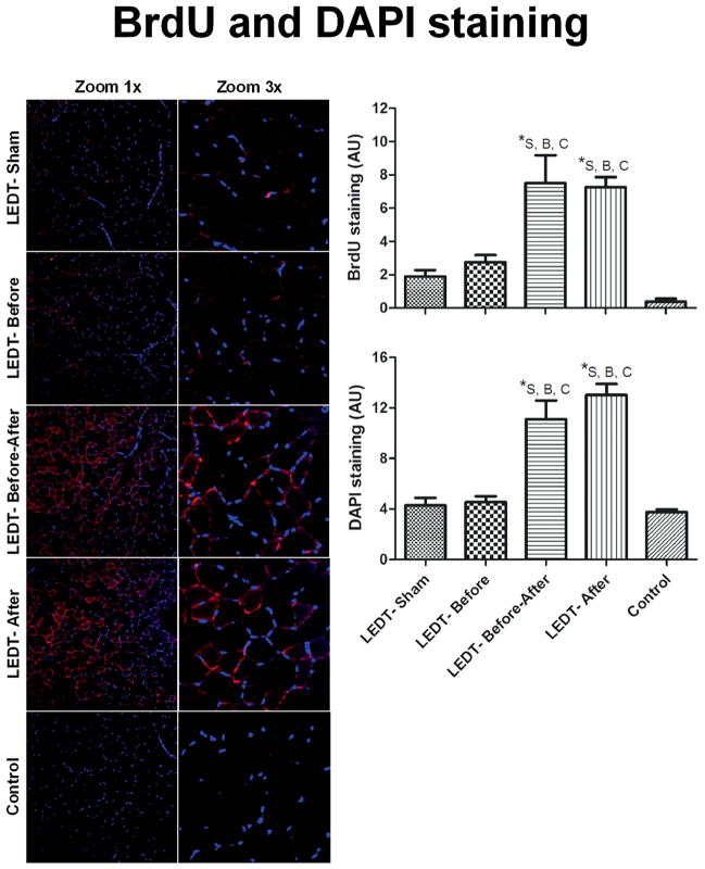
Figure 7.
Muscle cells in proliferative state and adult myonuclei in gastrocnemius muscle (n = 5 animals per each trained group; n = 2 animals in control group). BrdU (5-bromo-2′-deoxyuridine) immunofluorescence stained positive muscles cells in proliferative state. All the red staining indicates newly formed myonuclei. Purple dots mean the merge of red (new myonuclei in formation) and blue (adult myonuclei). DAPI (4′,6-diamidino-2-phenylindole) stained adult myonuclei or already formed myonuclei. Images were acquired with confocal microscopy (Olympus America Inc. Center Valley, PA, USA) at a magnification of 20× with zoom of 1× and 3×. BrdU and DAPI staining were quantified using software Image J (NIH, Bethesda, MD). * statistical significance (p < 0.05). Abbreviations: LEDT = light-emitting diode therapy; LEDT-Sham (Sham – S) = LEDT placebo (LEDT device in placebo mode) on muscles immediately before (5 minutes) each training session on ladder; LEDT-Before (Before – B) = LEDT applied on muscles immediately before (5 minutes) each training session on ladder; LEDT-Before-After (Before-After – A–B) = LEDT applied on muscles immediately before (5 minutes) and immediately after (5 minutes) each training session on ladder; LEDT-After (After – A) = LEDT applied on muscles immediately after (5 minutes) each training session on ladder. Control (C) = not submitted to any exercise or muscle performance assessment. AU = arbitrary units. Comparisons among all groups were conducted using One-way analysis of variance (ANOVA).

