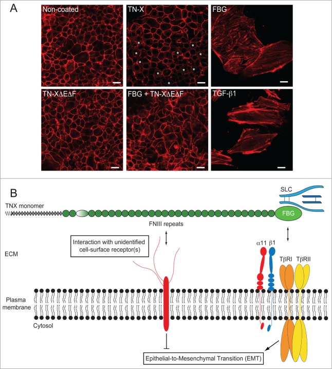
Figure 5.
The FNIII region of TN-X antagonizes the induction of EMT by the FBG-like domain. (A) Actin direct fluorescence in normal murine mammary epithelial (NMuMG) cells seeded onto non-coated dishes or dishes containing equimolar quantities of immobilized recombinant full-length TN-X, FBG-like domain, FNIII repeats (TN-XΔEΔF) or both FNIII repeats and FBG-like domain. Note that compared to the uncoated condition, where almost all cells exhibited a cortical actin staining, the full-length TN-X induced a mild EMT, visualized by a partial delocalization of actin cytoskeleton from cell junctions (*). In contrast, the FBG-like domain caused a full EMT, as illustrated by the acquisition of elongated cell morphology and the organization of actin cytoskeleton into stress fibers. Recombinant human TGF-β1, used here as a positive control, gave similar results to those obtained with the FBG-like domain. However, the FNIII region of TN-X fully inhibited the EMT triggered by the FBG-like domain. Bars, 15 μm. (B) Model representing the regulation of epithelial cell plasticity by TN-X. When separated from the intact TN-X molecule, the FBG-like domain is able to induce a robust EMT response, through its ability to activate latent TGF-β. This response relies on the presence of TβRII/I receptors and α11β1 integrin at the cell surface. In the intact TN-X molecule, the FNIII region antagonizes the EMT induced by the FBG-like domain, most likely via distinct intracellular cues that are initiated by the interaction of certain FNIII repeats to a yet unidentified receptor.

