
Fig. 6.
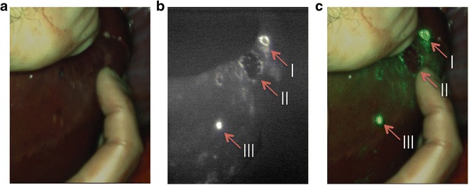
Near-infrared fluorescence imaging of colorectal liver metastases: 24 h after injection of 10 mg indocyanine green, colorectal liver metastases could clearly be identified by a rim around the tumor (I and II). Benign lesion (III) could be identified by fluorescence without the rim. Images are depicted in a visible light, b NIR fluorescence signal, and c a real-time overlay.