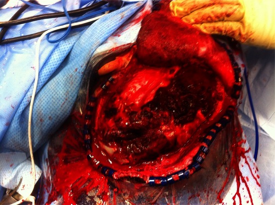
Fig. 1.

Operative photograph showing a gunshot wound to the left parietal lobe. The craniotomy has been performed and the dura opened. The upper part of the ear lobe is exposed in the upper operative field. Note the gross hemorrhagic track of the bullet with surrounding swollen brain. The wound was contaminated with dirt
