. 2015 Apr 30;19(3):219–228. doi: 10.4196/kjpp.2015.19.3.219

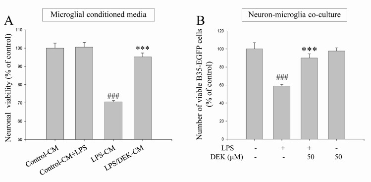
Fig. 2. Neuroprotective effects of DEK against microglial-mediated neuronal cell death. (A) BV-2 microglial cells were pretreated with DEK (50 µM) for 6 h. After wash-out, microglial cells were further incubated with LPS (1 µg/ml) for 24 h in the absence of DEK. MTT assay-based cell viabilities of HT-22 neurons were measured after different conditioned media treatment for 24 h. Groups are summarized as follows: the conditioned media from control BV-2 cells (Control-CM); LPS was added to the conditioned media from control BV-2 cells (Control CM+LPS); the conditioned media from LPS alone-treated BV-2 cells (LPS-CM); the conditioned media from LPS-treated BV-2 cells after DEK pretreatment (LPS/DEK-CM). (B) BV-2 microglial cells were pretreated with DEK (50 µM) for 6 h and then washed. B35-EGFP neurons were added to these BV-2 microglial cells to construct neuron-microglia co-culture system. Then, LPS (1 µg/ml) was stimulated for 24 h in the absence of DEK. The numbers of B35-EGFP neuronal cells were assessed by counting EGFP-positive cells under a fluorescent microscope. Fluorescent images of five random fields per well were captured and counted. Values are the mean±S.E.M. of four samples in one independent experiment. The data were replicated in three repeated independent experiments. ###p<0.001 as compared to the untreated control group and ***p<0.001 as compared to LPS alone-treated group.

Fig. 2