. 2015 Apr 30;19(3):219–228. doi: 10.4196/kjpp.2015.19.3.219

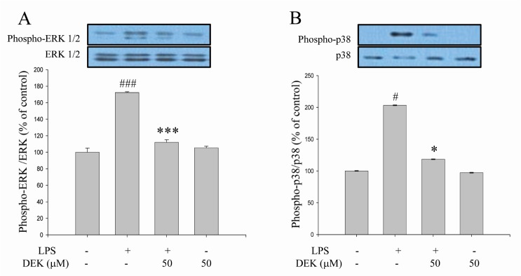
Fig. 3. Inhibitory effects of DEK on ERK phosphorylation in LPS-stimulated microglia. BV-2 microglial cells were pretreated with DEK (50 µM) for 1 h and then stimulated with LPS (1 µg/ml) in the presence of DEK for 30 min. Total proteins were subjected to SDS-PAGE, and probed with antibodies against phospho-ERK (A) and phospho-p38 (B) using Western blot analysis. Optical densities of individual protein bands were normalized to the corresponding levels of ERK and p38. Values are the mean±S.E.M. of four samples in one independent experiment. The data were replicated in three repeated independent experiments. #p<0.05, ###p<0.001 as compared to untreated control group and *p<0.05; ***p<0.001 as compared to LPS alone-treated group.

Fig. 3