
Figure 3.
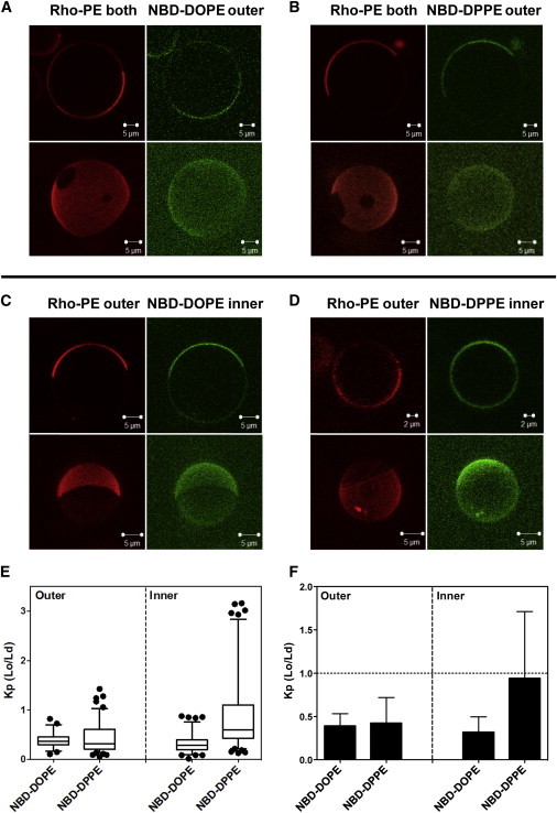
Images and NBD partition data for asymmetric egg SM + DOPCo/DOPCi/∼37 mol % cholesterol GUV. GUV labeled with (A) NBD-DOPE or (B) NBD-DPPE in the outer leaflet, and labeled with NBD-DOPE (C) or with NBD-DPPE (D) in the inner leaflet. (C and D) To restrict NBD fluorescence to the inner leaflet, outer leaflet NBD was reduced with sodium dithionite. For GUV with outer-leaflet NBD lipid, Rho-DOPE was in both leaflets; for GUV with inner-leaflet NBD, lipid Rho-DOPE was exchanged into the outer leaflet. Note that the vesicles in (D) are from the less abundant subpopulation in which NBD-DPPE preferentially associates with Lo domains. (A–D) Two-dimensional cross-section images (upper panel) and three-dimensional reconstruction images (bottom panel) are shown. Two- and three-dimensional images are from different vesicles. (E and F) Box plot and bar graph representations, respectively, of NBD-DOPE and NBD-DPPE Kp (Lo/Ld). Box boundaries at 25% and 75% values, whiskers set at 5% and 95% values. NBD-DOPE in the outer leaflets, n = 23; NBD-DOPE in the inner leaflet, n = 37; NBD-DPPE in the outer leaflet, n = 46; NBD-DPPE in the inner leaflet, n = 44. To see this figure in color, go online.