
Figure 3.
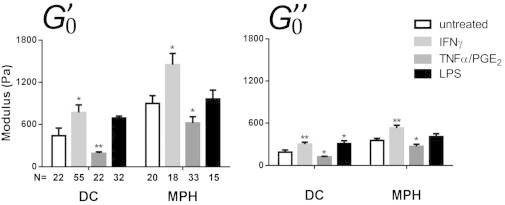
Storage () and loss () moduli of DCs and MPHs after treatment with different inflammation factors. Cells were incubated with IFNγ (light gray), TNFα/PGE2 (dark gray), or LPS (black) for 24 h before testing (∗p < 0.05, ∗∗p < 0.01, according to Mann-Whitney U test compared with untreated cells; N, number of cells tested, from at least three different donors). For values, see Table 1.