
Figure 3.
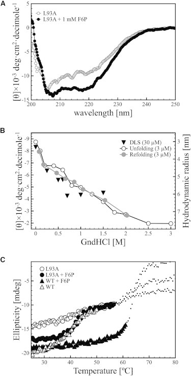
Structural features of the L93A mutant of Pfk-2 in the absence and presence of F6P. (A) Mean residue ellipticity of L93A Pfk-2 before (open circles) and after addition of F6P at a final concentration of 1 mM (black circles). (B) Change in the hydrodynamic radius (black triangles) and the mean-residue molar ellipticity of L93A Pfk-2 at a wavelength of 220 nm upon unfolding (open circles) and refolding (gray circles) as a function of the concentration of GndHCl. The protein concentration used was 3 μM for the CD measurements, and 30 μM for the DLS measurements. (C) Change in the ellipticity of WT Pfk-2 (triangles) and L93A (circles) upon increasing the temperature in the absence (open symbols) and presence (black symbols) of 1 mM F6P. The dotted lines represent the CD signal measured after the high-tension transition for each sample. The protein was incubated with F6P for at least 40 min at 20°C before the experiments.