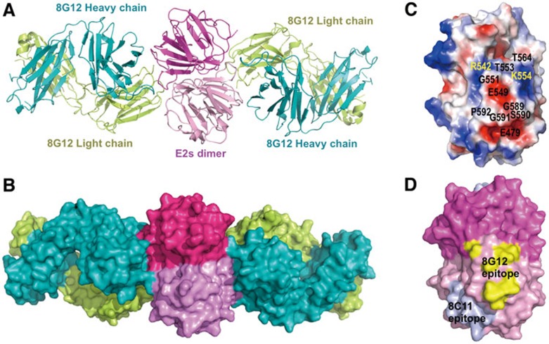
Figure 3.
Crystal structure of the Fab of cross-genotype neutralizing mAb 8G12 in complex with HEV capsid protein E2s domain. (A) Cartoon representation shows the dimeric E2sHEV-4:8G12 Fab complex. 8G12 heavy chain (H) and light chain (L) are depicted in cyan and yellow, respectively. In the dimeric E2s, one monomer is shown in dark pink and the other in light pink. (B) Surface representation for the E2sHEV-4:8G12 Fab complex. (C) Image depicts the electrostatic surface potential of the dimeric E2sHEV-4 (red, negative; blue, positive; gray, neutral) with the key residues of the 8G12's epitope are marked. The dimeric E2sHEV-4 is shown in an orientation in which most of the epitopes are visible. More epitopes are from one E2s monomer while only two residues (R542 and T564) from the other monomer (D) Surface representation shows the mapping of the epitopes of 8G12 and 8C11 on E2sHEV-1/4. Structure-related figures were prepared using the program PyMol37.

