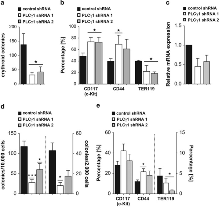
Figure 3.
Plcγ-1 regulates colony formation in erythroid progenitors and primary FLCs. (a) I/-11 cells infected with either Plcγ1 or control shRNA were plated in methylcellulose supplemented with Epo (10 U/ml) and transferrin (0.5 mg/ml); colonies were counted after 10 days. Each experiment was done in triplicate, error bars represent mean±S.E.M. (n=4). (b) Immunophenotype of colonies was investigated by flow cytometry using markers against CD44, TER119 and CD117 (c-Kit). Error bars represent mean±S.D. (n=4). (c) Quantitative RT-PCR of Plcγ1 mRNA in FLC after infection with either Plcγ1 shRNAs or control shRNA. Each experiment was done in triplicate and the error bars represent mean ± S.D. (n=2). (d) FLC of C57BL/6J mice (five independent experiments, n=5) were harvested at day E13.5 and infected with either Plcγ1 or control shRNA. For each experiment, cells were seeded in methylcellulose supplemented with cytokines at two different concentrations (10 000/2000 cells) and colonies were counted after 10 days. Each concentration in each independent experiment was done in triplicate, error bars represent mean±S.E.M. (e) Immunophenotype of colonies was investigated by flow cytometry. Error bars represent mean±S.E.M. (n=5)

