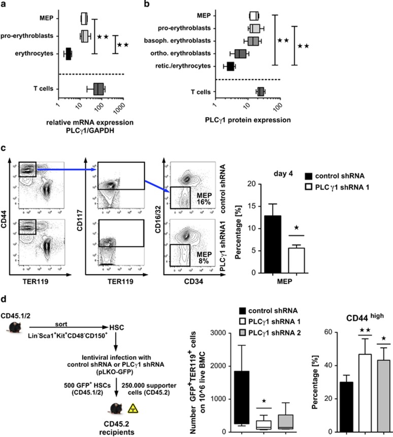
Figure 4.
Plcγ1 is essential for erythroid differentiation of adult BM cells in vitro and in vivo. (a) Relative expression of Plcγ1 mRNA in sorted BM cells of C57BL/6J mice (n=5), determined by qPCR. Each experiment was done in triplicate, error bars represent mean±S.D. (b) Intracellular Plcγ1 protein expression was analyzed in different BM compartments of C57BL/6J mice (n=5) using flow cytometry. Error bars represent mean±S.D. (c) Lineage-depleted/erythroid-enriched (Gr1-, B220-, CD3/4/8-, CD19-/IL-7Rα-negative) BM cells of C57BL/6J mice (n=4) were infected with either Plcγ1 or control shRNA. Differentiation was measured by flow cytometry over a time period of 96 h (day 4). A representative FACS blot (left panel) and percentage of MEP cells after 96 h (day 4; right panel) is shown. Error bars represent mean ±S.D. (d) Immunophenotypically defined HSC (Lin−Sca1+KIT+CD48−CD150+) were sorted and infected with Plcγ1 shRNA or control shRNA. 500 GFP+ HSCs were injected along with 2.5 × 10⁵ supporter cells (whole BM) into lethally irradiated recipient mice. Twenty weeks after transplant, the mice were killed and BM was evaluated for erythroid lineage development; for flow cytometry analysis cells were labeled with antibodies against TER119 and CD44. For each group, five mice were analyzed; error bars represent mean ±S.D. (n=5)

