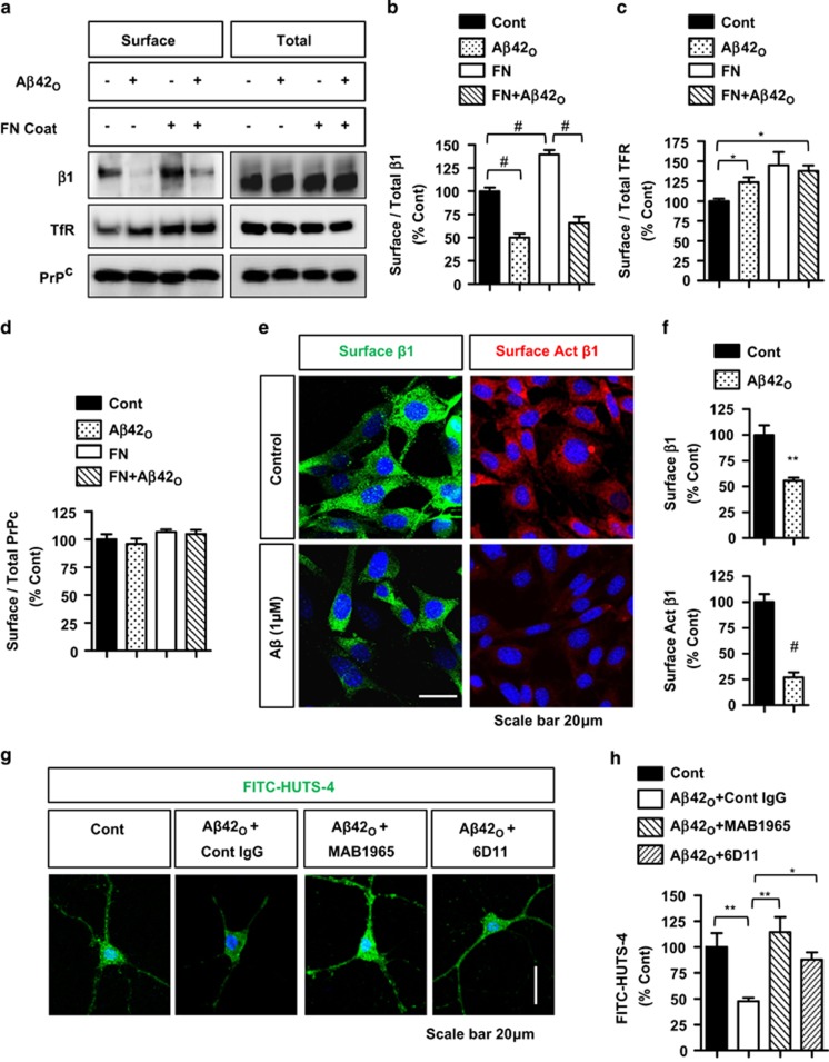
Figure 1.
Aβ42O induces loss of cell surface β1-integrin but not TfR or PrPc and reduces β1-integrin activation. (a–d) HT22 cells plated with/without FN, treated with/without Aβ42O (1 μM, 2 h), subjected to surface biotinylation (surface), and direct immunoblotting (total) or immunoprecipitation for biotin (surface) and immunoblotting for the indicated proteins. (b–d) Quantification of normalized surface/total β1, TfR, and PrPc (n⩾3 replicates, ANOVA, post hoc Tukey,*P<0.05, #P<0.0005). Error bar represent S.E.M. (e and f) HT22 cells treated with /without Aβ42O and immunostained for total surface β1-integrin or activated surface β1-integrin (HUTS-4 antibody) without membrane permeabilization (n⩾4 replicates, T-test, **P<0.005, #P<0.0005). (g and h) Direct staining for activated β1-integrin (FITC-HUTS-4 antibody) in fixed and permeabilized DIV14 primary hippocampal neurons after treatment with/without Aβ42O (1 μM, 2 h) and/or control ascites IgG (1 : 250), MAB1965 (1 : 250), or 6D11 (1 : 50). (h) Quantification of FITC-HUTS-4 immunoreactivity (n=4 replicates, ANOVA, post hoc Tukey,*P<0.05, **P<0.005)

