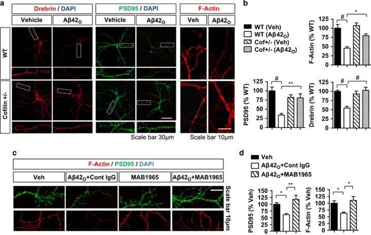
Figure 4.
Essential roles of Cofilin and β1-integrin in Aβ42O-induced depletion of F-actin and associated synaptic proteins in primary hippocampal neurons. (a and b) WT and Cofilin+/− DIV21 hippocampal neurons treated with/without Aβ42O (1 μM, 2 h) and stained for F-actin (rhodamine-phalloidin), Drebrin, or PSD95. (b) Quantification of mean intensities of Drebrin, PSD95, and F-actin in spine-containing neurites (n=4–6 replicates, ANOVA, post hoc Tukey, *P<0.05, **P<0.005, #P<0.0005). Error bars represent S.E.M. (c and d) DIV21 hippocampal neurons treated with/without Aβ42O (1 μM, 2 h) and/or control IgG or β1-integrin IgG (MAB1965) and stained for PSD95 and F-actin (rhodamine-phalloidin). (c) Representative images showing depletion of PSD95 and F-actin after Aβ42O treatment, which is prevented by MAB1965. (d) Quantification of mean PSD95 and F-actin intensities in spine-containing neurites (n⩾6 replicates, ANOVA, post hoc Tukey, *P<0.05, **P<0.005)

