Figure 5.
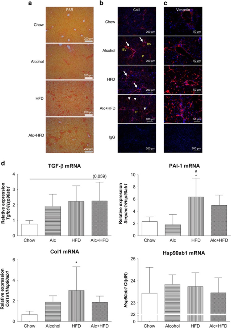
Alcohol and HFD induced hepatic fibrogenesis in mice. (a) Alcohol and HFD increased total collagen. PSR staining of paraffin-fixed liver sections showed increased collagen deposit in HFD- and Alc+HFD-treated mice compared with Chow. × 10 Magnification. (b) Alcohol and HFD increased Col1 (immunofluorescence). Immunofluorescent staining of frozen mouse liver tissues with anti-collagen 1 (Col1) antibody showed increasing collagen deposition (red) with all treatments around central and portal veins (arrows) with maximum staining visible in Alc+HFD mice livers across the lobule (arrowheads) compared with the other groups. Nuclei are stained with DAPI (blue). × 10 Magnification. (c) Alcohol and HFD increased vimentin-positive stellate cells in the liver. Vimentin-positive activated hepatic stellate cell (red) numbers increased with all treatments compared with Chow. Maximum increase was observed in HFD- and Alc+HFD-treated mice livers. Nuclei are stained with DAPI (blue). × 40 Magnification. IgG control at × 10 magnification. (d) Alcohol and HFD induced mRNAs of fibrosis-related molecules (Q-PCR). Expression of Col1 mRNA increased with all treatments compared with Chow but only HFD showed significance. TGF-β mRNA expression showed an increasing trend in all treatment groups compared with Chow but did not reach significance. PAI-1 mRNA expression increased significantly in HFD-treated mice compared with Chow and Alcohol groups. PAI-1 also increased with Alc+HFD but did not reach significance. mRNA expression of target genes was normalised using housekeeper Hsp90ab1. Hsp90ab1 mRNA expression (Ct) did not change between treatment groups (n=5–6). Data are mean±s.d.; *P<0.05 from Chow; #P<0.05 from Alcohol. BV, Blood vessel; P, Parenchyma; Ct, cycle threshold.

