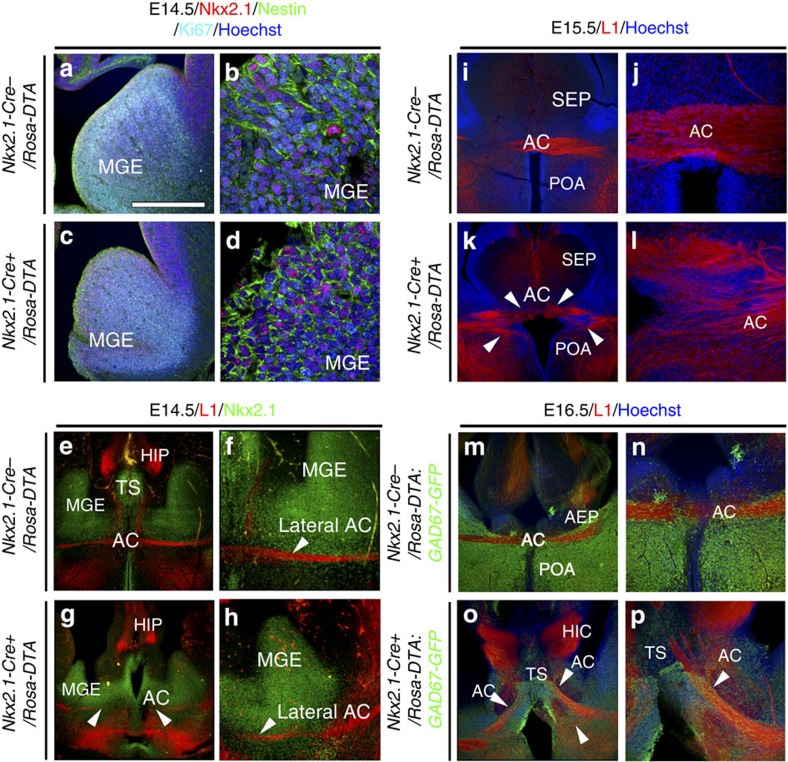
Figure 5. Early midline guidance defects of the AC in Nkx2.1-Cre+/Rosa-DTA mice.
(a–h) Triple immunochemistry for Nkx2.1, Ki67 and Nestin (n=4; a–d) and double immunochemistry for L1 and Nkx2.1 (n=3; e–h) on E14.5 coronal slices from Nkx2.1-Cre−/Rosa-DTA (a,b,e,f) and Nkx2.1-Cre+/Rosa-DTA (c,d,g,h) mice. (b,d) High-magnified views of MGE seen in a,c, respectively. (f,h) Higher power view of MGE and the lateral side of the AC seen in e,g, respectively. (a–d) Labelling with the cell cycle marker Ki67, combined with Nestin, revealed active proliferation of Nkx2.1+ progenitors in the MGE precursor regions in both control (a,b) and mutant (c,d) brains. (e–h) At E14.5, we could clearly see that in control (e,f) and mutant (g,h) brains, the L1-positive tracts behaved similarly within the lateral part of the AC. (i–l) Immunochemistry for L1 in coronal sections from control Nkx2.1-Cre−/Rosa-DTA (n=3; i,j) and Nkx2.1-Cre+/Rosa-DTA (n=3; k,l) mice at E15.5. (j,l) High-magnified views of the AC midline seen in i,k, respectively. (i,j) In Nkx2.1-Cre−/Rosa-DTA control mice, L1+ commissural axons crossed the AC midline and grew towards the contralateral cortex. (k,l) By contrast, in mutant Nkx2.1-Cre+/Rosa-DTA mice, defasciculation of axonal tracts at the AC midline can be seen and axons do not cross the midline (solid arrowheads). (m–p) Double immunochemistry for green fluorescent protein (GFP) and L1 on coronal sections from control Nkx2.1-Cre−/Rosa-DTA:GAD67-GFP (n=3; m,n) and Nkx2.1-Cre+/Rosa-DTA:GAD67-GFP (n=3; o,p) mice at E16.5. (n,p) High-magnified views of the AC midline seen in m,o, respectively. (o,p) In Nkx2.1-Cre+/Rosa-DTA:GAD67-GFP mice, there was a severe disorganization of GAD67-GFP+ interneurons. At E16.5, axons of the AC still did not cross the midline and are separated into two tracts that point ventrally and dorsally in the TS. HIC, hippocampal commissure; HIP, hippocampus. The scale bar shown in panel a corresponds to the following length for panel(s) specified in brackets: 675 μm (a,c,e,g,i,k,m,o); 320 μm (f,h,n,p); 160 μm (j,l); 50 μm (b,d).

