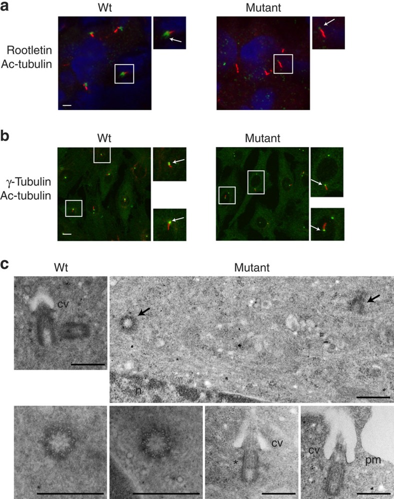
Figure 4. Ciliogenesis and ultrastructure of mutant centrioles observed using transmission electron microscopy.
(a) Ciliogenesis in vivo in adult kidney cells. Cilia were observed on kidney sections from wild-type and mutant cattle using acetylated tubulin to label the axoneme (in red) and rootletin (in green, arrow) to stain the cilium base. Enlargements show a cilium with strongly reduced rootletin staining in the mutant context. Distribution of cilia in SHGC mutant tissue was similar to the wild type, despite the very severe reduction of rootletin labelling (white arrow). Scale bar, 10 μm. (b) Ciliogenesis in fibroblast cells with split centrosomes. Cilia presence and length were analysed after labelling the axoneme with acetylated tubulin (in red) and the basal body with gamma-tubulin antibodies (in green, arrow). Enlargements of cells are shown to highlight the cilia. Mutant cells have the ability to grow and maintain cilia as efficiently as wild-type cells (see Supplementary Fig. 3 for statistics). Scale bar, 10 μm. (c) Normal centriole ultrastructure and cilium assembly in SHGC mutant fibroblasts. Wild-type centrioles were found as pairs in 50% of the sections analysed (n=20/42), while mutant centrioles were found isolated in 93% of the sections examined (n=91/98) or distantly located (black arrows). Transverse sections through the basal body shows the normal 9 microtubule triplets arrangement in the mutant centrioles and longitudinal sections allows the detection of subdistal appendages (*) and distal appendages that allow ciliary vesicle docking (cv), and subsequent fusion with the plasma membrane (pm). n, nucleus. Scale bar, 500 nm.

