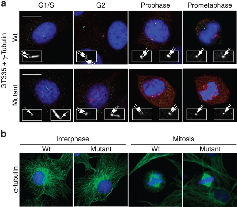
Figure 5. Bipolar mitotic spindle formation with paired centrioles in SHGC mutant cells.
Images show examples of microtubule, centrosome and centriole staining in wild-type and SHGC mutant primary fibroblasts at different stages of the cell cycle. (a) Newly formed centrioles remain associated with parental centrioles through the cell cycle in C-Nap1-deficient cells. Fibroblasts were synchronized in G1/S with thymidine and released for 8 h to observe cells in G2 or in various mitotic phases such as prophase and prometaphase. Centrioles and cilia are labelled with antiglutamylated tubulin monoclonal antibody GT335 (green), the centrosome with anti-gamma-tubulin serum (red), nuclei are stained with DAPI (blue). Insets are enlargements of GT335 labelling to highlight the number of centrioles (two centrioles in G1/S and four centrioles in G2 and mitosis). Note the presence of cilia in some G1/S and G2 cells. White arrows point each centriole. Merged images are stacks of four sections of 0.2 μm. Scale bar, 10 μm. (b) C-Nap1-deficient cells do not exhibit major defects in microtubule organization in interphase or mitosis. Asynchronously growing fibroblasts were immunolabelled for alpha-tubulin in green and DNA was stained with DAPI (blue). N=50 mitotic mutant cells were examined to assess spindle bipolarity.

