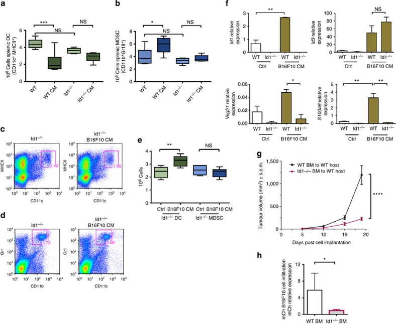
Figure 2. Deletion of the Id1 gene restores myeloid differentiation defects.
Flow cytometry analysis of spleens from WT and Id1−/− mice that received daily injections of B16F10 melanoma-derived conditioned media (B16F10 CM) or control media for (a) absolute numbers of DCs (one-way analysis of variance (ANOVA), ***P<0.001) and (b) absolute numbers of MDSC levels (one-way ANOVA, *P<0.05). (c) Representative frequency plots of DCs and (d) Splenic MDSCs isolated from Id1−/− mice injected daily with B16F10 melanoma-derived TCM or control media. (e) In vitro differentiation of Lin− haematopoietic progenitors isolated from Id1−/− mice, cultured for 6 days in the presence of B16F10 melanoma TCM (25% v/v) and analysed for DC and MDSC content by flow cytometry (n=6, ANOVA, **P<0.01). (f) Gene expression analysis of Id1−/− and WT cells after 6 days of in vitro differentiation in the presence of TCM, as determined by qPCR analysis (means±s.e.m., n=6, one-way ANOVA, **P<0.01, *P<0.05). (g) Analysis of primary tumour volume from Id1−/− and control BM chimeric mice following implantation of B16F10 melanoma cells (two-way ANOVA, ****P<0.0001). (h) Relative quantification of mCherry-labelled B16F10 melanoma cells in cryosections of lungs of Id1−/− and control BM chimeric mice measured by mCherry qPCR analysis (unpaired t-test, *P<0.05). NS, not significant.

