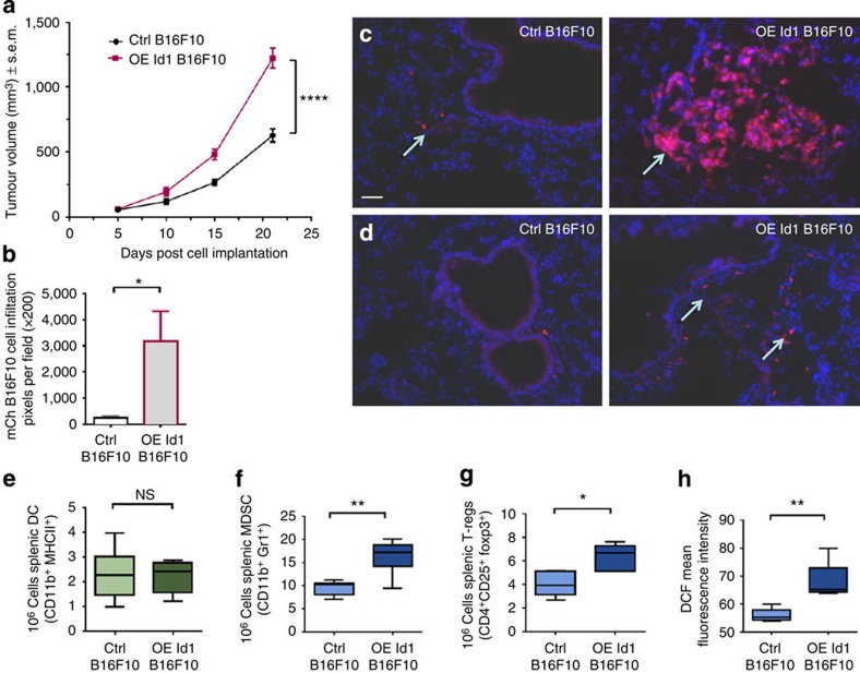
Figure 5. Id1-overexpressing BMDCs promote tumour growth and metastatic progression.
(a) Analysis of primary tumour volume from Id1-overexpressing mice and control vector mice following implantation of B16F10 melanoma cells (two-way analysis of variance, ****P<0.0001). (b) Quantification of mCherry-labelled B16F10 melanoma cells in cryosections of lungs of BM Id1-overexpressing mice and control vector mice measured as red pixels per field (unpaired t-test, *P<0.05). (c) Macro- and (d) micrometastatic lesion formation in lungs from Id1-overexpressing mice and control vector mice; scale bar (50 μm) on top left panel applies to all panels. Flow cytometry analysis of splenocytes from BM Id1-overexpressing and control vector mice implanted with B16F10 melanoma cells for absolute numbers of (e) DCs (f) MDSCs (g) regulatory T cell (T-regs) and (h) ROS production (unpaired t-tests; NS, non-significant, *P<0.05, **P<0.01). Four independent experiments were performed.

