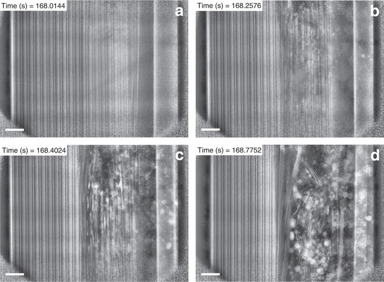
Figure 6. Radiographs from Supplementary Movie 5 showing the propagation of thermal runaway in Cell 1.
(a) Radiograph of the YZ plane before thermal runaway; (b,c,d) sequential images showing the propagation of thermal runaway through the cell. The thermal runaway initiates at the inner layers where the maximum temperature is apparent and spreads radially outwards. The formation of copper globules can be observed as highly attenuating white blots in images b, c and d. Heating is applied from the right of the images but continuous rotation at 180° every 0.4 s maintains an even circumferential temperature distribution. Scale bar, 1 mm.

