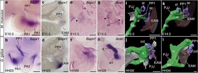
Figure 3. Comparison of development of the TM and PJJ in mouse and chicken.
(a–d) Bapx1 expression in pharyngeal arches of the embryonic day 10.5 mouse (a), stage 23 chicken (b) in lateral view and transverse sections of the embryonic day 11.5 mouse (c) and stage 25 chicken (d). Bapx1 and Aggrecan (Acan) expression in sagittal serial sections of embryonic day 12.5 mouse (e,g) and stage 26 chicken (f,h). The expression signals in the incus (i) and malleus (m) are indicated by arrowheads (e,g). (i–l) Three-dimensional reconstruction of forming the pharyngeal skeleton and TM of embryonic day 12.5 (i) and 14.5 (k) mouse, and stage 26 (j) and 34 (l) chicken (coloured same as in Fig. 1). art, articular; col, columella auris; EAM, external auditory meatus; Mc, Meckel's cartilage; PA1–2, pharyngeal arches 1–2; PJJ, primary jaw joint; PP1, first pharyngeal pouch; q, quadrate; sp, styloid process; st, stapes. Scale bars, 200 μm.

