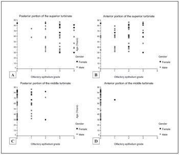
Figures 2A, 2B, 2C, 2D. 2.

A: Light micrograph (400x magnification) of a grade 0 anterior middle turbinate sample. This slide contains only respiratory epithelium; note the predominance of goblet cells. 2B: Light micrograph of a posterior superior turbinate fragment with a predominance of olfactory neuroepithelium. Note the isolated focus of respiratory epithelium between the arrows. This specimen was graded as 3 + . 2C: Light micrograph of a posterior superior turbinate fragment, lower magnification. Olfactory neuroepithelium predominates in this specimen; note the thin basal membrane and cellular lamina propria. 2D: Light micrograph of a posterior middle turbinate fragment, 200x magnification. This specimen contains mostly respiratory epithelium. Note the thick basal membrane and predominance of vascular structures in the lamina propria.
