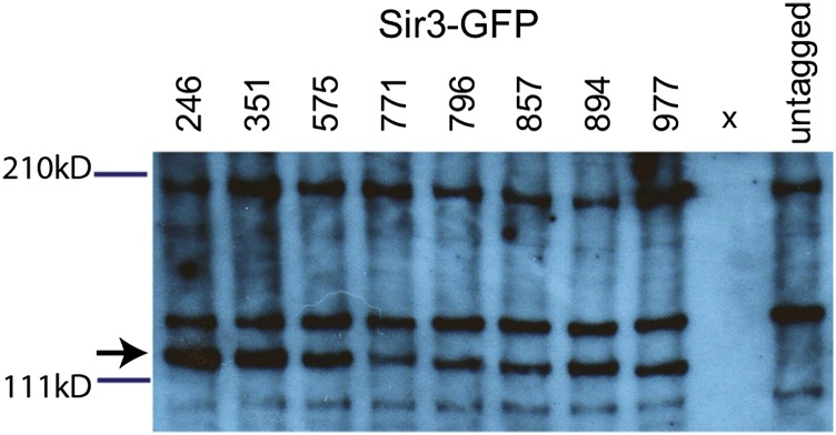
Figure 7.

Western blot analysis of Sir3-GFP in C. glabrata. An anti-GFP immunoblot of C. glabrata strains carrying different Sir3-GFP alleles. Wells are labeled with the amino acid site of the GFP insertion within Sir3; “x” indicates an empty lane. The antibody cross-reacted with C. glabrata whole-cell lysates, as shown by the banding see in the untagged strain. The band representing Sir3-GFP is marked with an arrow.
