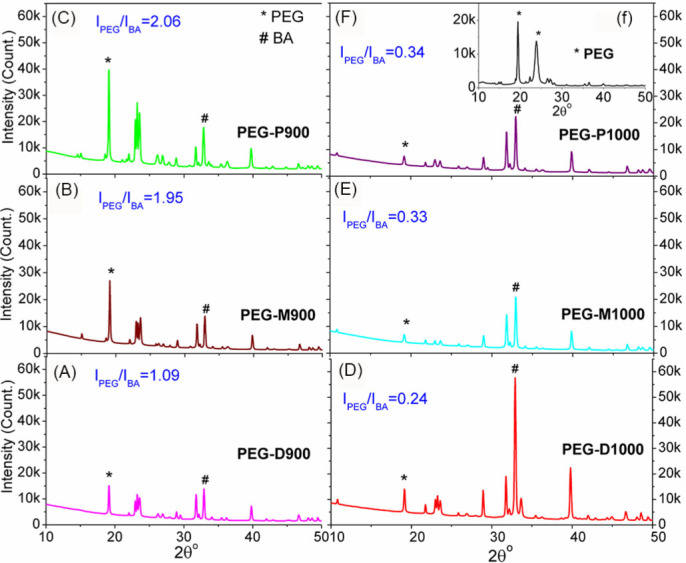
Figure 2. XRD patterns of the PEG-impregnated bovine apatite (BA) scaffolds: (A) PEG-D900, (B) PEG-M9000, (C) PEG-P900, (D) PEG-D1000, (E) PEG-M1000, and (F) PEG-P1000; inset (f) XRD pattern of pristine PEG.

Intensity ratio (IPEG/IBA) of PEG (IPEG) to BA (IBA) correspond to their maximum intensities (semicrystalline peak of PEG ‘*’ and crystalline peak of BA‘#’) indicates the quantitative presence of PEG in BA.
