Figure 3.
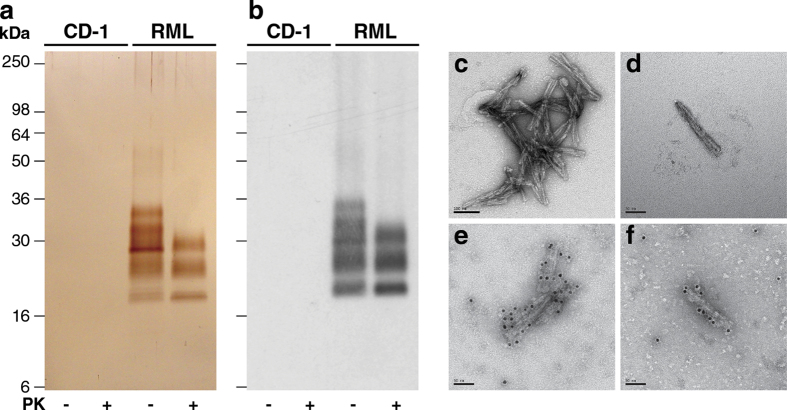
Purified RML prions from CD1 mouse brain. (a,b) P4 fractions from uninfected (CD1) or RML-infected (RML) mouse brain purified with (+) or without (–) PK-digestion. (a) Silver-stained 16% SDS-PAGE gel. (b) Western blot using anti-PrP monoclonal antibody ICSM35. The equivalent of 200 μl (a) or 20 μl (b) of 10% (w/v) brain homogenate was loaded per lane. (c-f) Electron microscopy images of prion rods in P4 fractions from RML-infected CD1 mouse brain obtained without PK digestion. Samples were stained with uranyl acetate. Prion rods in panels e and f have been labelled with anti-PrP monoclonal antibody SAF-32 and an anti-mouse IgG secondary antibody conjugated to 10 nm gold particles. Scale bar, (c) 100 nm, (d–f) 50 nm.

