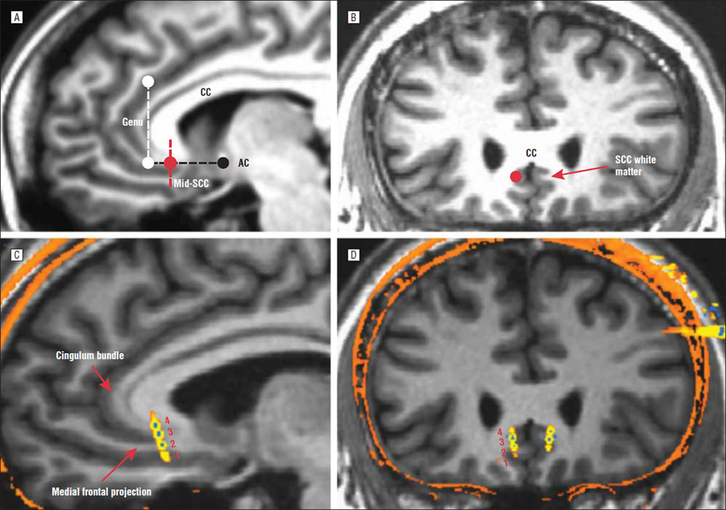
Figure 1.
Surgical targeting. Preoperative magnetic resonance imaging (MRI) shows the sagittal (A) and coronal (B) views of the planned optimal subcallosal cingulate (SCC) white matter target (red circle). The dotted black line indicates the subcallosal plane of interest, parallel to the anterior-posterior commissural line; the dotted white line indicates the rostral limit of the subcallosal plane; and the dotted red line indicates the midsubcallosal plane. The red circle indicates demarcation of the SCC white matter target and surrounding gray matter (best seen in the coronal view [B]). C and D, Postoperative computed tomography scan merged with preoperative MRI showing a typical case with the deep brain stimulation electrodes in situ. Note that the contacts span the SCC gray and white matter in the vertical plane proximal to the split of the cingulum bundle and rostral medial frontal white matter tracts (C, red arrows, sagittal view). Contacts are numbered by convention (1–4 on the left, 5–8 on the right), inferior to superior. Contacts 2 and 3 are directly in the SCC white matter, and contacts 1 and 4 are in the inferior and superior gray matter, respectively. AC indicates anterior commissure; CC, corpus callosum.

