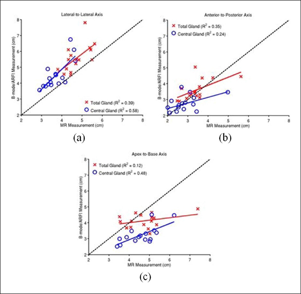
Figure 9.
Measurements of the prostate dimensions along the three standard anatomic axes: lateral-tolateral (a), anterior-to-posterior (b), and apex-to-base (c). The correlation between the MR and B-mode/ARFI imaging axis measurements was performed in each orientation for the total gland (red crosses) and central gland (blue circles). B-mode images were used for total gland measurements, and ARFI images were for central gland measurements. The black dashed-line represents the projection of perfectly correlated measurements between imaging and pathology. The over-/under-estimation of each imaging modality relative to gross pathology and each other is summarized in Table 2. MR = magnetic resonance; ARFI = Acoustic Radiation Force Impulse.

