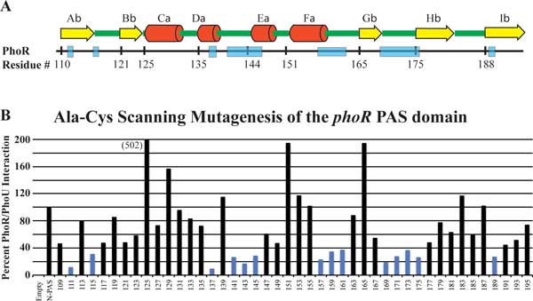
Figure 6.

PAS domain scanning mutagenesis. A. Predicted secondary structure of the PhoR PAS domain with alpha helixes labeled with yellow arrows and β-sheets labeled with red cylinders. B. Scanning mutagenesis of the PhoR PAS domain used BACTH to identify regions of the protein that are essential for interaction with PhoU. Every two amino acids were changed to code for alanine and cysteine. Each construct was tested in triplicate for interaction with PhoU. Blue bars represent samples that had less than 40% of the activity of unmutated PhoR.
