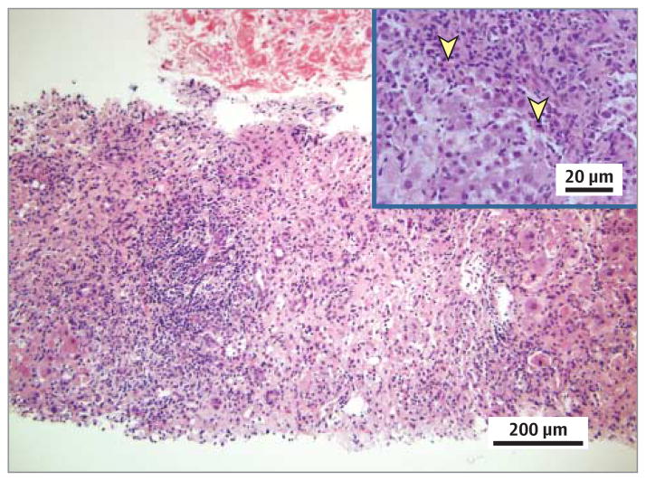
Figure 2. Results of the Second Liver Biopsy.
Lobular necrosis and portal and interface inflammation with predominant lymphocytes and scattered eosinophils (yellow arrowheads) are seen (hematoxylin-eosin, original magnification ×200). Inset is taken from a portion of the biopsy not captured on the larger slide (original magnification ×400).

