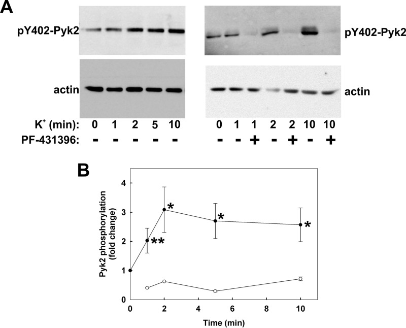
FIGURE 4.
Effect of K+-induced depolarization on Pyk2 autophosphorylation and its sensitivity to PF-431396. Rat caudal arterial smooth muscle strips were stimulated at time 0 with K+ following pre-incubation for 30 min with vehicle or PF-431396 (10 μm). Tissues were quick-frozen at the indicated times for analysis of Pyk2 autophosphorylation at Tyr-402 by SDS-PAGE and Western blotting with anti-pTyr-402-Pyk2. Loading levels were normalized to actin. A, representative Western blots for phosphorylated Tyr-402 Pyk2 (pY402-Pyk2) and actin. B, cumulative quantitative data expressed as -fold change relative to untreated tissue and normalized to actin. Values represent the mean ± S.E. (n = 13 for solid circles and n = 5 for open circles). *, p < 0.01, **, p < 0.02, significantly different from Pyk2 phosphorylation level prior to membrane depolarization.

