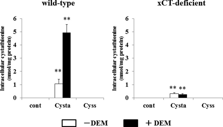
FIGURE 8.
Intracellular cystathionine levels in MEF derived from WT and xCT-deficient (KO) mice. WT and KO MEF were cultured for 24 h in the routine culture medium, and then cultured further for 24 h with or without 50 μm diethyl maleate (DEM). Then the cells were incubated for 15 min in 1 ml of PBS(+)G (PBS containing 0.1% glucose, 0.01% Ca2+, and 0.01% Mg2+) with or without 0.1 mm cystathionine or 0.1 mm cystine. After a 15-min incubation, intracellular cystathionine levels were determined as described under “Experimental Procedures.” Each point represents the mean ± S.D. (error bars) (n = 3–8). **, p < 0.01 compared with control.

