FIGURE 3.
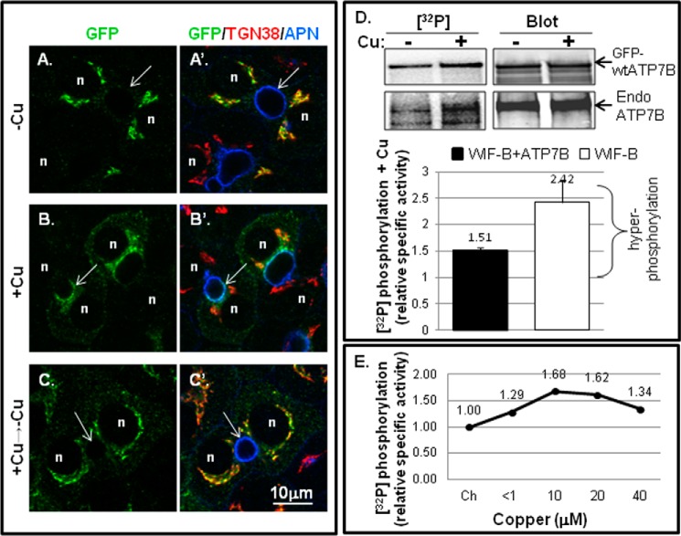
Copper-stimulated ATP7B trafficking and Ser/Thr phosphorylation in WIF-B cells. WIF-B cells were incubated overnight in 10 μm BCS (−Cu) to stage endogenous ATP7B or GFP-WTATP7B in the TGN. For the +copper (+Cu) condition, cells were incubated in 10–20 μm CuCl2 for 1–1.5 h. A–C′, WIF-B cells infected with GFP-WTATP7B were fixed; stained with anti-GFP (green), TGN38 (red), and aminopeptidase N (APN) (blue); and imaged by confocal microscopy. Single plane images show copper-dependent trafficking. n, nucleus; arrows, apical surface. D, representative autoradiogram of 32P incorporation and corresponding immunoblot of immunoprecipitated endogenous (GFP-wtATP7B; top gel bands) and (Endo ATP7B; bottom gel bands) from metabolically labeled WIF-B cells ±copper (top). The bar graph (bottom) shows the mean relative specific activity normalized to chelator-treated cells. n = 7 for GFP-ATP7B (black bar; left); n = 3 for endogenous ATP7B (white bar; right). Error bars represent S.E. E, dose response of hyperphosphorylation to varying copper concentrations. <1, the copper level in basal medium. The relative specific activity of 32P-labeled GFP-WTATP7B in WIF-B cells is shown normalized to the value obtained in chelator (Ch).

