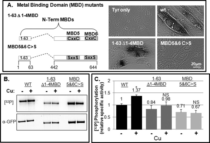
FIGURE 4.

N-term MBD mutants exhibit basal phosphorylation and not copper-dependent hyperphosphorylation. A, schematic showing N-terminal MBD mutants (right). YST cells were co-transfected with pTyrosinase alone (Tyr) or with various ATP7B constructs as indicated. The black reaction product indicates the presence of copper-dependent tyrosinase activity (left). B and C, GFP-WTATP7B and the two N-terminal mutants were expressed and evaluated for phosphorylation as described under “Experimental Procedures.” B, autoradiogram and blot showing 32P incorporation and protein levels of the three constructs. C, bar graph showing the mean relative specific phosphorylation of GFP-WTATP7B, 1–63 Δ1–4MBD, and 1–63 Δ1–4MBD C>S normalized to the −copper (−Cu) condition. n = 4. Error bars, mean ± S.E.; *, p < 0.05 using two-tailed t test; NS, not significant.
